Programmed death-ligand 1 (PD-L1), also well-known as B7-H1 and CD274, is a protein that acts as a kind of 'brake' to keep the body's immune responses under control. PD-L1 may be found on some normal cells and in higher-than-normal amounts on some types of cancer cells. When PD-L1 binds to programmed death-1 (PD-1, also known as CD279 and PDCD1), a protein found on T cells, it keeps T cells from killing the PD-L1-containing cells, including the cancer cells. Anticancer drugs called immune checkpoint inhibitors bind to PD-L1 and block its binding to PD-1. This releases the 'brakes' on the immune system and leaves T cells free to kill cancer cells.
| Localization | Function | Tissue specificity |
|
|
|
PD-1 is involved in immune tolerance by suppressing activated immune cells via interaction with its ligands. Two known ligands of PD-1 are PD-L1 and PD-L2. PD-L1 is a 290 aa transmembrane glycoprotein. PD-L2 may lead to local cytokine production that is beneficial to the tumor cells. PD-L1 and PD-L2 play different roles in the immune regulatory process. PD-L1 inhibits T-cell function in peripheral tissues, whereas PD-L2 suppresses immune T-cell activation in lymphoid organs.
The PD-1/PD-L1 pathway represents an adaptive immune resistance mechanism exerted by tumor cells in response to endogenous immune anti-tumor activity. PD-L1 is overexpressed on tumor cells or non-transformed cells in the tumor microenvironment. The binding of PD-L1 to PD-1 leads to the inhibition of the cytotoxic T cells. These deactivated T cells remain inhibited in the tumor microenvironment.
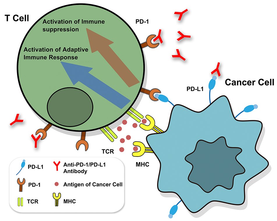 Fig.1. Identification of PD-1/PD-L1.1,2
The monoclonal antibody (mAb) therapies against PD-1 and PD-L1 are routinely used for immunotherapy. The efficiency of the immune checkpoint blockade with mAbs in cancer treatment is remarkable, but not all patients respond to a single therapy. To enhance and broaden the anti-tumor activity of immune checkpoint inhibition, one possible solution is combinational immunotherapy. An example of this is the success of the combination of PD-1/PD-L1 inhibition blockage with complementary checkpoint inhibitor CTLA-4 in melanoma and non-small cell lung cancer.
Creative Biolabs offers a wide range of services designed to study the immune checkpoints, including blocking antibodies, proteins, assays, and more. Our experienced service team can also save your time by developing reagents to meet your specifications. We provide the following services for worldwide customers.
For any questions, please feel free to contact us for more information.
References
All listed customized services & products are for research use only, not intended for pharmaceutical, diagnostic, therapeutic, or any in vivo human use.
Our customer service representatives are available 24 hours a day, 7 days a week.
USA
Tel:
Fax:
Email:
Copyright © 2026 Creative Biolabs. All Rights Reserved.
