Recently, there was a publication by a research team. They found that Lymphocyte Activation Gene-3 (LAG-3) in neurons is a specific receptor for Tau Pre-formed Fibril (PFF) in the brain, which promotes the propagation of Tau PFF between neurons. In addition, they observed that if LAG-3 was knocked down in neurons, it significantly reduced the endocytosis of Tau PFF and decreased its propagation between neurons. This result also implies that LAG-3 may serve as a potential therapeutic target for AD and Tau protein disorders.
Creative Biolabs offers a wide range of services designed to study immune checkpoints, including LAG-3. Our experienced service team provides the following services for worldwide customers. Please do not hesitate to contact us if you are also conducting research on the immune checkpoint LAG-3.
| Services | What We Do | Advantages |
| LAG-3 Immune Checkpoint Molecule for Drug Development | We provide customized protein development, antibody development, targeted peptide development, targeted small molecule drug development for the immune checkpoint LAG-3, immune checkpoint assays, and preclinical studies for LAG-3-targeted drugs. |
|
| Immune Checkpoint Binding and Interaction Assays | We can offer a comprehensive set of binding and interaction assays to screen for promising candidates. With our customized project design, we can demonstrate your molecules' binding, interaction, and functional properties. |
|
| Immune Checkpoint Antibody Development | Our comprehensive portfolio of services includes polyclonal antibody discovery and development, monoclonal antibody discovery and development, antibody fragment discovery and development, antibody characterization, antibody engineering and optimization, and antibody production and manufacturing. Targets include but are not limited to, PD-1, CTLA-4, LAG-3, TIM-3, TIGIT and BTLA. |
|
Protein aggregation is a major pathological feature of degenerative diseases such as Alzheimer's disease (AD) and Parkinson's disease (PD). In AD and PD, for example, tau proteins and α-synuclein are altered in the pathological state, and further aggregated to form a propagating "seed" (e.g., PFF, which is also an aggregate of tau), which can enter the cell to induce intracellularly derived protein misfolding and aggregation and propagate between neurons by intercellular transmission, causing protein aggregates to persist to fibrillar and insoluble fiber morphologies. Interneuron propagation, which sustains the protein aggregates towards the morphology of PFF and insoluble fibers, and eventually protein deposits and tangles.
There have also been studies identifying some receptors associated with tau protein endocytosis and spreading, such as acetylheparan sulfate proteoglycan and low-density lipoprotein receptor-associated protein 1. However, these receptors are not specific for tau PFF.
Some researchers discovered back in 2016 that a cell surface receptor, LAG-3, can bind specifically to the PFF of α-synuclein, and that inhibiting LAG-3 inhibits the uptake of α-synuclein and reduces pathological aggregation of α-synuclein. On this basis, the researchers wondered whether LAG-3 could also bind specifically to tau PFF.
In the latest study, the researchers first extracted AD-tau from the brains of AD patients, then obtained tau PFF using sonication, and investigated the specific binding of LAG-3 to tau PFF by a series of checkpoint assays.
In addition to its direct interaction with tau protein aggregates, emerging evidence suggests that LAG-3 may play a crucial role in the intercellular transmission of pathological tau species between neurons. Tau propagation, the process by which misfolded tau proteins spread from one neuron to another, is believed to contribute to the progressive spread of tau pathology throughout the brain in tauopathies. Recent studies have implicated LAG-3 in mediating tau neuron-to-neuron transmission, providing further insight into the pathophysiological mechanisms underlying disease progression.
To observe the role of LAG-3 in tau PFF interneuron propagation, the researchers performed a microfluidic device to simulate interneuron propagation. The results revealed that LAG-3 plays a key role in tau PFF interneuron propagation.
From these results, it is clear that LAG-3 can bind specifically to tau PFF and promote the propagation of tau PFF between neurons, so LAG-3 may serve as a potential therapeutic target for tau protein disorders.
LAG-3 as a Therapeutic Target for AD
The identification of LAG-3 as a key player in tau pathology has positioned this immune checkpoint molecule as a promising therapeutic target for AD and related tauopathies. Targeted modulation of LAG-3 activity holds the potential to intervene at multiple stages of tau-mediated neurodegeneration, from the initial aggregation of tau proteins to the propagation of pathological tau species throughout the brain.
After injecting LAG-3 antibodies into cortical neurons, researchers found that LAG-3 antibodies not only significantly blocked the binding of LAG-3 to tau PFF, but also reduced the transmission of tau PFF between neurons.
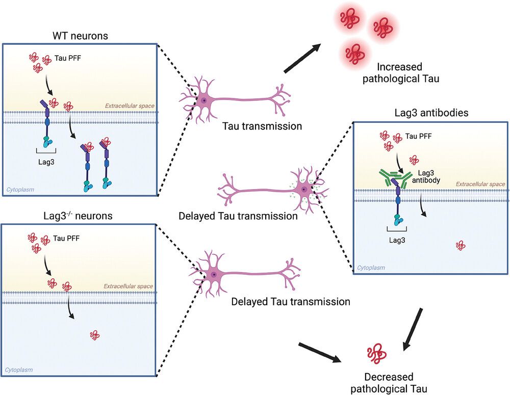 Fig. 1 Binding of LAG-3 to LAG-3 antibody significantly reduces Tau PFF binding and endocytosis, leading to delayed propagation of pathology and toxicity.1
Creative Biolabs is at the forefront of LAG-3-targeted therapeutics development, leveraging cutting-edge technologies and innovative approaches to harness the therapeutic potential of LAG-3 modulation in neurodegenerative diseases.
Reference
All listed customized services & products are for research use only, not intended for pharmaceutical, diagnostic, therapeutic, or any in vivo human use.
Our customer service representatives are available 24 hours a day, 7 days a week.
USA
Tel:
Fax:
Email:
Copyright © 2024 Creative Biolabs. All Rights Reserved.